
Pyoderma gangrenosum on a residual limb: when the wound gets worse with pressure
A September 2026 O&P EDGE article describes a clinician's first encounter with pyoderma gangrenosum in a prosthetic patient. The condition is rare, often misdiagnosed as infection, and has a defining feature that makes it particularly difficult in an amputee context: it gets worse when you apply the kind of mechanical trauma that prosthetic socket management usually involves.
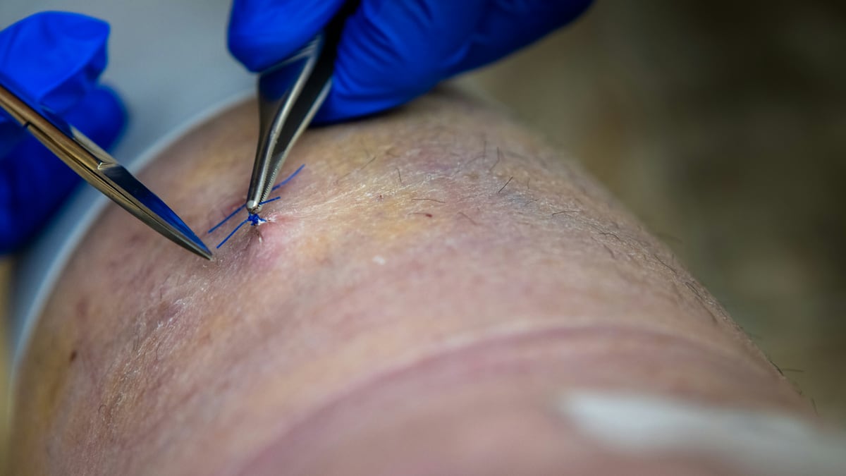
The O&P EDGE published an article this month from a clinician who had not, until encountering one specific patient, heard of pyoderma gangrenosum. The piece describes the experience of working through an unfamiliar diagnosis and adapting socket management accordingly. It is written for practitioners. What it points to, for patients and families, is worth explaining in plain terms: there is a skin condition that looks like an infected wound, is often treated as one, and gets measurably worse when clinicians apply the interventions that would normally be correct.
Pyoderma gangrenosum is rare. Estimates put its incidence at roughly three to ten people per million per year, and there is no reason to think amputees face a substantially elevated baseline risk. But when it appears on a residual limb, its clinical profile collides directly with the mechanics of prosthetic use in ways that are worth understanding if you or someone on your care team encounters it.
What the condition is
Pyoderma gangrenosum is a neutrophilic dermatosis. That means it is driven by an abnormal accumulation of neutrophils, the immune cells that normally respond to bacterial infection, in tissue where no infection is causing the response. The skin ulcers that result are painful, often violaceous at the border, and can progress from a small nodule or pustule to a large open wound in days.
It is associated with underlying systemic conditions in roughly half to three-quarters of cases. Inflammatory bowel disease, both Crohn’s and ulcerative colitis, is the most common association. Rheumatoid arthritis and hematologic conditions including myeloid leukemia and myeloma are also associated. In a significant minority of people, no underlying condition is found: the pyoderma is the primary diagnosis.
There is no definitive diagnostic test. Diagnosis is clinical, involving the wound’s appearance, its course, a biopsy to rule out other causes, and consideration of whether an associated systemic condition is present. Because the presentation can look like a severe skin infection or a vascular ulcer, misdiagnosis is common and has direct consequences for treatment.
The defining feature: pathergy
The property that makes pyoderma gangrenosum particularly dangerous to mismanage is pathergy. Pathergy means the wound worsens in response to mechanical trauma.
In practice, this includes debridement, which is normally a standard component of wound care for ulcers. It includes biopsy. And it includes the sustained pressure of a prosthetic socket against the wound site or surrounding skin. Rather than improving after these interventions, a pyoderma wound tends to enlarge. Surgical debridement of a pyoderma lesion, done because the wound looks necrotic or infected, often dramatically worsens the wound. There are documented cases of a small ulcer expanding to cover a large surface area following what appeared to be appropriate wound care.
This is why getting the diagnosis right is not merely procedural. Treating pyoderma gangrenosum as an infected socket sore, and responding with the tools typically used for infected socket sores, can cause the wound to grow faster than if it were left alone with topical antibiotics and reduced socket contact.
What treatment looks like
Once pyoderma gangrenosum is identified, treatment targets the underlying immune dysregulation rather than any infection. Topical corticosteroids or tacrolimus ointment may be appropriate for small or early lesions. More extensive wounds typically require systemic therapy: high-dose oral corticosteroids are the most common initial approach, with cyclosporine and biologic agents including infliximab used in refractory or corticosteroid-dependent cases.
Wound care is handled gently. Aggressive debridement is avoided. Dressings aim to protect the wound surface from mechanical disruption rather than to remove tissue.
For prosthetic users, this means the interaction between wound management and socket use has to be managed explicitly by the care team. A wound on the residual limb may require a period without socket use while systemic treatment is initiated. Return to prosthetic use, once the wound is responding, involves careful interface management, attention to pressure distribution, and monitoring for any sign that socket contact is worsening the healing trajectory.
What this means for residual limb wound management
Most wounds on a residual limb are mechanical in origin: pressure sores, shear injuries, folliculitis, fungal overgrowth. These are common in prosthetic users, they have well-understood causes, and they respond to established interventions. Pyoderma gangrenosum is uncommon enough that most amputees will never encounter it and most O&P clinicians will see it rarely or not at all.
The O&P EDGE article is worth noting precisely because the practitioner who wrote it had not seen it before. That is normal. It is a rare condition.
What the rarity does not mean is that a wound which looks wrong should be assumed to be ordinary. A residual limb wound that is unusually painful relative to its size, that appeared rapidly, that is progressing despite appropriate socket modifications and wound care, or that is worsening rather than improving following debridement, is a wound that warrants dermatology involvement. The practitioner in the O&P EDGE article eventually brought in dermatology and worked from there. That collaboration is the model: socket design adaptations are part of the management, but they follow from getting the diagnosis right, not the other way around.
If you are managing a wound on your residual limb that is not responding as expected, the relevant question to bring to your care team is whether the wound has had dermatology assessment. The O&P clinician alone is not the right arbiter of a wound that may have an inflammatory rather than mechanical origin.
This desk covers clinical information for context. Nothing here is medical advice or a treatment recommendation for a specific situation. Wound management on a residual limb should involve your medical and O&P care team.
Source notebook: This reporting draws on The O&P EDGE: Choosing the Right Socket for Pyoderma, September 2026 ↗. We link out so you can follow the receipts.